Tensor Fasciae Latae Syndrome and Lateral Entrapment with Instability
Nancy Martin-Molina, DC, QME, MBA, CCSP
Case History
FK is a 67-year-old woman with a six-month history of right-sided hip pain. She attributed the pain onset to crawling on her side under her home patio deck to replace dry rot and consequently felt she might have twisted something in her right hip. On initial presentation, she reported that the pain was worse with physical activity, described as "prolonged walking." She also experienced difficulty moving from a seated to a stance position, or in any up-step activity. She reported relief once in a stance position. She denied any urologic or neurologic symptoms. There was no loss of bowel, bladder or sexual function. She was retired and normally enjoyed an active lifestyle that included gardening and walking three miles a day, at least three to four days a week.
The patient's prior medical history is significant for status post four-year right-sided abrasion chrondoplasty involving the right knee, to which she reported full recovery. She had previously been seen for one month by another chiropractor without relief, and was without current medications at time of presenting with hip pain.
Examination revealed a youthful-appearing geriatric woman complaining of moderate pain (5 on a 1-10 visual analogue scale, or VAS), in the right greater trochanteric region (according to pain drawing). Physical examination was negative for hip pathology. She could forward flex to 70¯ without pain and exhibited a positive Minor's sign on arising, with complaints of localized ache of the affected hip. The patient was without paresthesias or hyporeflexia to indicate any specific nerve root involvement. Radiographic imaging of the spine and pelvis was felt to be the initial diagnostic test of choice to rule out neurologic or osseous disease.
Range of motion of FK's hips and joints in the lower extremities did not reproduce her pain. The distal vascular exam was normal. She presented with a functional right short leg. Palpable trigger point bands, purposeful withdraw and referred pain was demonstrated on digital examination of the tensor fasciae latae, and to a lesser extent in the gluteus maximus and quadratus lumborum. Ober's test revealed contracture of the right fascia lata. Tests for dynamic lateral entrapment were essentially negative (Pheasant test). Truck rotation in stance with pelvis held firmly was inconclusive at reproducing hip pain. Chiropractic assessment yielded joint fixation of L1-L2 vertebrae and pelvis. Postural examination revealed an antalgic stance and right toe-out posture.
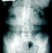
Radiographic weightbearing imaging studies were performed to evaluate the patient's lumbar spine and hips. There was marked reduction in disc height, retrospondylolisthesis of the first lumbar vertebra onto the second lumbar vertebra with reduction in size of foramen. There were proliferative osteophytes (overgrowth of bone forming hard bars) and moderate anterior traction spurring demonstrated. There was right laterolisthesis of the spinous processes accompanied by right concavity scoliosis. The acetabuli and hip joint spaces were well-maintained.
Discussion 1: Myofascial pain syndrome (a hyperirritable spot within a taut band of skeletal muscle or fascia).
Figure 1
Figure 2
Figure 3
Figures 1-3: Three images of patient FK's spine display the ravages of tensor fasciae latae syndrome, including reduction of disc height and foramen, spurring, and scoliosis. Figures 2-3 exhibit spurring.
This case initially illustrated the typical clinical findings of myofascial pain syndrome (MPS) presenting as chronic hip pain. Although the patient did have L1-L2 degenerative disc disease, she was essentially without low back pain. The major difficulties were related to the chronic hip pain disorder and its interference on her recreational activities described as walking. The MPS was identified as the tensor fascia latae muscle, which is part of the muscular component of the floor of the femoral triangle. Identification was made through reproduction of chief complaint on digital examination and clinical correlation.
Clinical Features 1: MPS
There are several clinical features of MPS due to trigger points (TPs) that warrant discussion. The exquisite local tenderness of the TP is well explained by sensitization of the nerve endings of group III and group IV muscle nociceptors. Substances that are known to affect sensitive tissues include bradykinins, prostaglandins, histamine and leukotrienes. There is a palpable band that is characteristic of myofascial pain and helps localize the involved muscles. The rope-like band produced on digital examination of the involved muscle fibers at the TP can be explained by contracture. Clinically, the patient will experience pain whenever tension is placed on the taut band muscle fibers. There is a localized shortening of a group of muscle fibers combined with cutting off local circulation of the capillaries in the TP zone strong enough to produce localized tissue hypoxemia (inadequate tissue perfusion).
MPS is a condition that is treatable by eliminating the specific trigger points that are the immediate cause of pain, and correcting those factors that predispose to recurrence. The diagnosis can be made by treatment of the affected region by myofascial release. Many chiropractic practitioners, often at pre-adjustment, utilize digital ischemic compression of the TP for 15-30 seconds, with rhythmic percussion of the TP at about two-second intervals to provide a counter-irritant to disrupt the TP.
Discussion 2: Lateral entrapment with instability (nerve entrapment in lateral part of nerve canal that runs from the medial edge of the superior facet to the foramen with instability defined as posterior facetal capsule laxity permitting joint subluxation in presence of internal disc disruption).
This case also illustrated the atypical clinical findings of lateral entrapment with instability presenting as chronic hip pain. Although the patient did have myofascial pain syndrome, the initial presenting mechanism of trauma (despite symptoms being in a chronic phase of care) did present as a shearing, twisting event. The major difficulties were then related to the co-morbidity of the spinal instability.
Clinical Features 2: Lateral Entrapment with Instability
The ways that a foramen may be diminished in size are basically threefold: loss in disc height, subluxation of facets, and stenosis, or narrowing of the intervertebral foramen. In the presence of instability, the lax facet capsule allows the superior facet to move backward and forward with rotation. In this patient, the spinous process of L1 and L2 have been rotated; the superior facet moved anteriorly; the joint space had opened and the lateral canal was markedly narrowed. In the presence of fixations or advanced degenerative changes to stabilize the affected segment, little or no movement should take place at affected level. The lateral canal is fixed because of the degenerative deformity. Thus, two factors may be involved in producing the entrapment: subluxation of posterior facets, (superior facet moves superior and anterior), and enlargement of facet by osteophytic involvement that further narrows the lateral canal. The diagnostic imaging of choice is a CT scan.
FK was treated 20 times over eight weeks under my direction with good response. Conservative chiropractic treatment included high velocity, low force lumbar and pelvic adjustments. Pre-adjustment therapy included: Percussor therapy, ultrasound with trigger point head, and myofascial release with digital ischemic pressure. Post adjustive therapy included tensor fascia latae stretches. Active muscle balancing exercises were introduced. Follow-up visits were arranged once every two weeks over the next three months as she returned to her walking activity.
Here is a classical example of approaching the patient goals threefold: Foremost, always address the patient's chief complaint and correct it before addressing any other complaints (find the primary reason for the visit), obtain a primary diagnosis and a differential diagnosis, format a treatment plan that address each condition. Too often patients come into my practice after having seen other colleagues, and when asked why they did not continue care, the responses are generally: "I really didn't feel they addressed my complaint," or "They kept trying to adjust my neck; that wasn't my problem."
Stay focused and continue to promote chiropractic. Eventually patients will understand why their necks may need adjusting.
References
Mense S. Nervous outflow from skeletal muscle following chemical noxious stimulation. J Physiol 267:75-88, 1977.
Travell JG, Simmons DG. Myofascial Pain and Dysfunction: The Trigger Point Manual. Williams & Wilkins, 1992.
Gerwin RD. The management of myofascial pain syndromes. Journal of Musculoskeletal Pain (The Haworth Press, Inc) Vol. I, No. 314, 1993, pp. 83-94.
Simmons DG. Myofascial pain due to trigger points. International Rehabilitation Medicine Association, IRMA Monograph Series Number 1, Nov 1987.
Travell JG, Simmons DG. Myofascial origins of low back pain. Postgraduate Medicine, Low Back Pain, Part I, Vol. 73/No 2/ February 1983.
Rubin D. Myofascial trigger point syndromes: an approach to management, Arch Phys Rehabil Vol. 62, March 1981.
Travell JG, Rinzler SH. The myofascial genesis of pain. Postgraduate Medicine Vol. II, No. 5, May 1952.
Kraus H. Evaluation and treatment of muscle function in athletic injury. Amer Journal of Surg, Vol. 98, September 1959.
Many relevant diagnostic signs are not performed deliberately by the examiner or by the patient at the examiner’s direction. They are observed as the patient reacts to their condition. Fortin’s finger sign, Minor’s sign, and Vanzetti’s sign are three examples of this principle.
Spearheaded by burgeoning scientific and clinical research literature, psychedelics have reached a level of media coverage and popular interest that has not been seen for over half a century. By “psychedelics,” we are referring to the unique class of substances that includes psilocybin (the active compound found in so-called “magic mushrooms”), LSD, dimethyltryptamine (DMT), ayahuasca, 5-MeO-DMT, and mescaline – each of which occurs in the natural world (except for LSD, which is a semi-synthetic compound).
While working at the Lovelace Medical Center in Albuquerque, N.M., one of the primary care physicians stated that imaging every patient prior to receiving chiropractic was not medically necessary. It was at that point I reconsidered the need for the imaging of every patient to avoid malpractice.