Patients may occasionally ask you to take a chest x-ray. Often the chest x-ray is a pre-employment requirement. If you choose to take chest x-rays for your patient, please be aware of the technical requirements. The accepted standard technique for taking a PA chest is non-grid at 72 inches, 100 to 110kVp, film size 14 x 17 in full inspiration. The central ray is approximately at T6. It is important to include the apices of the lungs and costophenic margins. The lateral chest is taken with the same technique, except for the positioning. The central ray on the lateral view should be in the axillary region. With a good technique the thoracic spine should be only slightly visualized on the PA view. The broncovasular markings should be clearly visualized, if they are not, you have over-penetrated. Normally the heart shadow is less than half the greater diameter of the thorax. If any finding appears suspicious, get a second opinion. I don't need to even discuss with you the malpractice implications if a neoplasm or even an infection is missed. If your patient is a smoker, I would recommend getting a second opinion, even if the study appears normal.
PA Chest:
Lateral Chest:
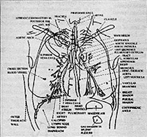
The Cardiothoracic Ratio:
This measurement is performed to determine the size of the heart and a screening for heart enlargement.
MRD = maximum transverse diameter of the right side of the heart, which is a line drawn from the midline of the spine to the most distant point of the right cardiac margin.
ML= midline of the spine
MLD = maximum transverse diameter of the left side of the heart, which is a line drawn from the midline of the spine to the most distant point of the left cardiac margin
Many relevant diagnostic signs are not performed deliberately by the examiner or by the patient at the examiner’s direction. They are observed as the patient reacts to their condition. Fortin’s finger sign, Minor’s sign, and Vanzetti’s sign are three examples of this principle.
Spearheaded by burgeoning scientific and clinical research literature, psychedelics have reached a level of media coverage and popular interest that has not been seen for over half a century. By “psychedelics,” we are referring to the unique class of substances that includes psilocybin (the active compound found in so-called “magic mushrooms”), LSD, dimethyltryptamine (DMT), ayahuasca, 5-MeO-DMT, and mescaline – each of which occurs in the natural world (except for LSD, which is a semi-synthetic compound).
While working at the Lovelace Medical Center in Albuquerque, N.M., one of the primary care physicians stated that imaging every patient prior to receiving chiropractic was not medically necessary. It was at that point I reconsidered the need for the imaging of every patient to avoid malpractice.