It is important to determine if anterior knee pain is from the local area or referred from a more proximal site. While pain could be referred to the anterior knee from the hip or spine, distal extremity complaints that can be palpated as local pain are most likely to be the site of the lesion, rather than being referred. Most patients entering our offices, especially athletes, have more involvement of the patellofemoral area than the intra-articular tibiofemoral joint. The largest anterior structure of the knee is the patella, and most of the time, the anterior knee pain is related to the patellar articulation or the surrounding tissues.
While there are of course many possible causes of anterior knee pain, such as patellar tendinosis, quadriceps tendinopathy, coronary ligament fibrosis, and prepatellar bursitis, to name a few, a particular passive structure that supports the patellar and knee has been recognized as a major source of pain. It is the patellar retinaculum. According to Biedert and Sanchis-Alfonso,1 free nerve endings, which besides detecting pain, heat and cold, also detect crude touch and pressure, have the highest density in the tendon of the quadriceps muscle and the second highest in the retinacular and patellar tendon. They feel that this is necessary since these tissues control acceleration, deceleration and rotation of the knee joint, and require high proprioceptive capability.
The retinacular, an important stabilizer of the patella, is composed of fibrous bands that form discrete ligaments attaching medially and laterally into the patella. Both sides of the retinacular help to keep the patellar in alignment. For example, as the knee flexes, the lateral retinacular bands that originate from the iliotibial band exert a normal posterolateral force on the patella. If the medial support of the patella becomes weakened over time, the lateral retinacular pressure will cause a lateral tilting of the patella, or if allowed to continue, eventual subluxation and even dislocation.
According to what is known as the "Law of Valgus," the lateral side of the knee, composed of the distal vastus lateralis, iliotibial tract and lateral retinaculum, is normally stronger and more fibrous than the medial forces, composed primarily of the distal vastus medialis obliquus (VMO) and the medial retinaculum. This explains why lateral compression syndromes develop in the knee, and why lateral release operations are often performed. A tight lateral retinaculum that tilts the patella causes increased pressure on the lateral facet and eventual adaptive changes in the patellar articular cartilage.
Kasim and Fulkerson2 evaluated the patella retinaculum in 25 patients, 20 of whom had experienced failed surgery such as realignment of the extensor mechanism or a lateral release procedure, and continued to complain of postsurgical anterior knee pain. The 25 patients with anterior knee pain underwent retinacular excisions followed by muscle flexibility and strengthening. Twenty-two of the 25 patients (88 percent) noted moderate to substantial improvement.
The most prominent cause of anterior knee pain has been blamed on what is called patellar malalignment, and surgical techniques are used to realign the patella. Kasim and Fulkerson stated that the cause of malalignment and patellofemoral pain often involves excess pressure on part of the patella due to imbalance of the retinacular restraints, causing the malalignment. Therefore, if we can find restrictive knee retinaculum, it is important to treat it and determine if the malalignment and patella tracking improves - and more importantly, whether the patient's symptoms improve. In their study, the authors found specific painful areas of the retinaculum that they excised. Histological studies revealed that the painful retinaculum contained neuromatous degeneration of the small nerves of the painful tissue and degenerated fascia, with no inflammation. It sort of resembled tendinosis-type tissue.
An important evaluation for a tight lateral restraint is use of the patellar tilt and glide test. The medial side of the patella can be tilted inferiorly to open the lateral side, at which point palpation of the lateral side would reveal tenderness. The lateral patella could be pushed (glided) medially, and failure of the patella to displace medially more than 1 cm would indicate a tight lateral retinaculum. The patella articulation (cartilage) would be suspected if the examiner compresses the patella and gets a painful response when the patient is asked to contract the quadriceps.
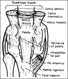
Treatment of choice is the Graston technique (see image left), since the instruments can be used to penetrate and release the retinaculum. The practitioner could open up the lateral side by pressing down on the medial side of the patella to reach more areas (not shown). Friction massage can also be attempted. All of the peripatellar retinaculum should be evaluated for tenderness and restriction. It is necessary to treat and evaluate the retinaculum with the patient supine and the knee extended, to allow freer motion of the patella while it is out of the trochlea sulcus. Stretching and strengthening of the related areas should also be part of the procedure.
References
Biedert RM, Sanchis-Alfonso V. Sources of anterior knee pain. Clin Sports Med 2002;21:335-347.
Kasim N, Fulkerson JP. Resection of clinically localized segments of painful retinaculum in the treatment of selected patients with anterior knee pain. Am J Sports Med 2000;28(6):811-814.
Spearheaded by burgeoning scientific and clinical research literature, psychedelics have reached a level of media coverage and popular interest that has not been seen for over half a century. By “psychedelics,” we are referring to the unique class of substances that includes psilocybin (the active compound found in so-called “magic mushrooms”), LSD, dimethyltryptamine (DMT), ayahuasca, 5-MeO-DMT, and mescaline – each of which occurs in the natural world (except for LSD, which is a semi-synthetic compound).
At its March 11, 2024, meeting, the Council for Higher Education Accreditation (CHEA) Committee on Recognition reviewed the formal request for a change in the CHEA-recognized scope of accreditation submitted by the Council on Chiropractic Education (CCE). The new CHEA-approved scope statement is: “The Council on Chiropractic Education accredits doctor of chiropractic degree programs and chiropractic residency programs in the United States, its territories and Canada (2024).”
Many relevant diagnostic signs are not performed deliberately by the examiner or by the patient at the examiner’s direction. They are observed as the patient reacts to their condition. Fortin’s finger sign, Minor’s sign, and Vanzetti’s sign are three examples of this principle.