It seems as if we don't order CT scans anymore - but don't sell this diagnostic tool short. Let me give you a brief overview of spiral computerized axial tomography (spiral CT).
It is relatively new, developed in the late 1980s and introduced in 1989 to the medical profession at the Annual Meeting of the Radiological Society of North America (RSNA). I say "relatively" new, because it has been in use for over 10 years; but magnetic resonance (MR) imaging has been so popular, and still is often the imaging modality of choice for musculoskeletal disorders, that we don't usually order CT scans much anymore, unless an MR is not available.
CT Explained
Don't worry - this is not a physics lesson! Spiral CT was made possible by the development of slip-ring technology. This is what allows the electrical energy to be transmitted to the gantry as it passes along the patient, so the acquisition of CT data is performed in a helical or spiral fashion. (Picture a corkscrew.) This allows for the anatomical data to be acquired as a volume, rather than as a series of slices. With previous CT scans, the gantry was only able to rotate once through 180/360 degrees before the scanner had to be reset for another scan to be performed, and the data was acquired in only one plane at a time, as a series of slices.
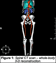
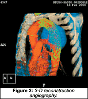
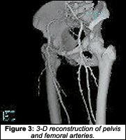
With spiral CT, the data only need be acquired once; since it is volume data, it can be segmented to give any desired slice thickness, and at any point along the slice volume. The other great feature is that spiral CT is fast - on the order of 20-60 seconds, which allows for the reduction or complete elimination of respiratory or any other motion artifacts. Even severely ill patients can hold their breath and stay still for 20 seconds. Moreover, because of the volume data and lack of motion, artifact image reconstruction can be extremely high-quality (i.e., 3-D images), and can be used for angiography.
However, there is a trade-off (besides the significant difference in how the high voltage is supplied to the slip-ring between manufacturers, which entails a separate discussion). I will tell you, though, that as you might suspect, spiral CT places an extremely high load on the X-ray tube and generator, since it is operating continuously as it acquires the data along the patient as a volume. This also requires a sophisticated cooling system. I don't know what the tube life for one of these spiral CT scanners is, but I wouldn't run out and buy a used one! That is all I am going to discuss about the technical aspect of this modality.
What about clinical uses? An initial review of the literature to date indicates spiral CT angiography is quite useful for evaluating the intracranial/carotid arteries; the aorta; the pulmonary arteries; the renal/splanchnic arteries; and the peripheral veins. The scan time for the patient is 30-60 seconds. Trauma patients benefit most from spiral CT and its rapid data acquisition, as they may have difficulty remaining still or assuming certain radiography positions. With spiral CT, areas that are difficult to evaluate, including the shoulder joint, pelvis, and spine, can be examined rapidly. Spiral CT scans also are lower in cost and are more easily available than standard CT.
Musculoskeletal spiral CT provides excellent multiplanar and 3-D images that allow for the important localizing information that can define a mass's shape and size, tumor margins and their effect on fascial planes. Detection of impeding fractures and the relationship of tumor-affected bone to weight-bearing areas can be evaluated with 3-D reconstruction.
Regarding spinal trauma: Multipla-nar imaging and 3-D reconstruction have a wide variety of application, including the detection of fracture, subluxation (yes, the medical literature acknowledges the term), locked facets and foreign bodies. There are many more applications, and this technology is still evolving - so check it out!
Once again, I thank Philips Medical Systems (www4.medical.philips.com:80/Education/education.asp) for permission to use its images.
Spearheaded by burgeoning scientific and clinical research literature, psychedelics have reached a level of media coverage and popular interest that has not been seen for over half a century. By “psychedelics,” we are referring to the unique class of substances that includes psilocybin (the active compound found in so-called “magic mushrooms”), LSD, dimethyltryptamine (DMT), ayahuasca, 5-MeO-DMT, and mescaline – each of which occurs in the natural world (except for LSD, which is a semi-synthetic compound).
At its March 11, 2024, meeting, the Council for Higher Education Accreditation (CHEA) Committee on Recognition reviewed the formal request for a change in the CHEA-recognized scope of accreditation submitted by the Council on Chiropractic Education (CCE). The new CHEA-approved scope statement is: “The Council on Chiropractic Education accredits doctor of chiropractic degree programs and chiropractic residency programs in the United States, its territories and Canada (2024).”
Many relevant diagnostic signs are not performed deliberately by the examiner or by the patient at the examiner’s direction. They are observed as the patient reacts to their condition. Fortin’s finger sign, Minor’s sign, and Vanzetti’s sign are three examples of this principle.