Most of Your Back Patients Need Specific Stabilizing Exercises
Warren Hammer, MS, DC, DABCO
Editor's note: This article was printed in the 6-17 issue, but was not printed in its entirety. Our apologies to Dr. Hammer and our readers.
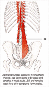
If you are not aware of the value of checking the lumbar muscles of stabilization, please read on. A major source of frustration after an acute low back pain (LBP) patient is discharged from care is the eventual recurrence of the problem. Hides, et al.1 proved the value of using specific stabilizing exercises to reduce the rate of recurrence of LBP. A principal lumbar stabilizer, the multifidus, has been found to be weak and atrophic in most acute LBP, and remains weak long after symptoms have abated.
The researchers divided 39 acute low back patients into a control group who received medical management and advice to resume normal activity as tolerated; another group performed specific localized exercises to restore the stabilizing protective function of the lumbar multifidus (LM) with co-contraction of the transverses abdominus muscle (TrA). All were given four weeks of exercise. The groups were followed by one and three-year questionnaires. Two to three years after treatment, the rate of recurrence of LBP in the specific exercise group recurrence was 35 percent, but 75 percent for the control group. The multifidi no longer showed weakness or atrophy in the specific exercise group.
Another study2 conducted a randomized, controlled trial on patients with chronic LBP and a radiologic diagnosis of spondylolysis or spondylolisthesis. At the 30-month follow-up, the specific exercise group showed a statistically significant reduction in pain intensity and functional disability levels.
There is a distinct difference between global and stabilizing muscles. Global muscles influence more than one joint or region, e.g., the rectus femoris influences the hip and knee; the thoracic erector spinae expand from the thoracic to the lumbar, ilium and sacrum. Stabilizing muscles are monoarticular muscles, such as the vastus medialis obliquus of the knee, which, instead of moving the knee, supports the patella during knee flexion and extension. Specific co-contraction of the TrA and deep portions of the LM do not produce spinal movement.3 The lumbar multifidi are more concerned with stabilization and control than spinal movement.
The TrA is another important stabilizing muscle, which, due to its transverse orientation, cannot contribute to extension, flexion or lateral flexion of the spine.3 These deep muscles close to the joint support the joint movement and increase the necessary joint stiffness of the lumbar spine. The TrA has a separate control system allowing independent action from the other trunk muscles. By way of the thoracolumbar fascia, it stabilizes the lumbar vertebrae by the fascia's attachment to the transverse and spinous processes. The LM that is contained within the posterior fascial sheath upon contracting also contributes to the tension of the posterior layer and affects lumbar stability. It has been found that the TrA contracts to stabilize the spine even before a person moves an arm or leg.4 In LBP patients, the onset of activity of the TrA is delayed with limb movement in all directions.3
I will attempt to explain how to check for weakness of the TrA and LM muscles, yet there is much more to realize to optimally evaluate these muscles and treat them. I highly recommend the textbook by Richardson, Jull, et al.,3 from which most of the information in this article is derived.
For the TrA, the weakness is most evident when the supine patient is asked to draw in the abdominal wall. The patient will usually use the external global muscles, especially the obliquus externus abdominus, and less often the obliquus internus abdominus, rectus abdominus and thoracic portions of the erector spinae. Often, these muscles are overactive, and are seen to react when the patient attempts to contract the TrA. They must be taught to contract the TrA in isolation from the other abdominal muscles. The patient must be told to concentrate on the lower part of the abdomen, especially since the lower portion of the TrA may be the most important part of the muscle, and is essential for spinal stabilization.3 The global abdominal muscles do not attach to the spine, but to the pelvis and rib cage. When attempting to draw the lower abdomen in, the patient should not move the spine or pelvis or take a deep breath. It is important to isolate the contraction of the TrA with "precision," rather than use great effort to tighten it.
Since the TrA contracts prior to other abdominal muscles (to stabilize the spine in preparation for movement by the global muscles), it is more important that the stabilizers contract rapidly than with additional strength. The patient and doctor can palpate the TrA slightly lateral and inferior to the anterior superior spine. It is a learning process to contract the abdomen below the umbilicus without contracting the abdominal muscles above the umbilicus, but it can be done. Statements such as "slowly draw in your lower abdomen away from the elastic of your pants," or "slowly draw your navel up and in toward your backbone"3 are helpful.
Weakness of the LM can be evaluated by having the prone patient extend one hip (10 degrees) at a time. The appearance of lumbar extension (lordosis), spinal rotation or lateral deviation are indicators of possible LM weakness.
While the patient is prone, the patient can become aware of the LM by the doctor sinking fingers deeply into the LM next to the spinous processes. Ask the patient to gently expand or contract his muscles against the fingers. A rapid or superficial contraction may indicate contraction of the longer superficial fibers of the multifidus, or tension in the tendinous portion of the thoracic erector spinae."3 A better facilitation of the contraction is occurring if there is a feeling of a deep and slowly generated tension under the fingers as the muscle expands against resistance.
A typical beginning exercise for the patient is to contract the TrA and hold it while in a crunch position for 10 seconds, and repeat 10 times. Patients can use this contraction in many types of spinal exercises, even when walking or driving. Because of co-contraction, every time they contract the TrA, they also will be contracting the LM; eventually patients will have their own protective brace. I explain to my patients that when they move their spines, especially if they suddenly reach for something, it is necessary that the spinal joints and capsules are not overly moving and stressing themselves, i.e., moving out of the stable "neutral zone," which Panjabi, et al., proved to be larger with intersegmental injury and intervertebral disc degeneration.5
The latest issue of Spine6 proved that the TrA also significantly decreased the laxity of the sacroiliac joints. It states: "The contraction of the TrA, independently of the other abdominal muscles, affects the laxity of the sacroiliac joints to a larger extent than a bracing action using all of the lateral abdominal muscles, and that exercises for lower back pain should incorporate a precisely controlled contraction of the TrA independently of the global muscles."
I have been teaching my patients lumbar stability exercises for several years and am convinced that the exercises reduce recurrent back pain. It is necessary to continually monitor patients, over time, to be certain that they are using the motor skills necessary to contract their TrA. Usually, when a patient returns with that old lumbar spasm, it is evident that he or she did not perform the exercises correctly, if at all.
References
Hides JA, Jull GA, Richardson CA. Long-term effects of specific stabilizing exercises for first episode low back pain. Spine 2001;26(11):e243-e248.
O'Sullivan PB, Twomey LT, Allison GT. Evaluation of specific stabilization exercise in the treatment of chronic low back pain with radiologic diagnosis of spondylolysis or spondylolisthesis. Spine 1997;24:2959-2967.
Richardson C, Jull G, Hodges P, Hides J. Therapeutic Exercise for Spinal Segmental Stabilization in Low Back Pain. New York, Churchill Livingstone, 1999:80-81.
Hodges PW, Richardson CA. Contraction of the abdominal muscles associated with movement of the lower limb. Phys Ther 1997;77:132-144.
Panjabi M, Abumi K, Duranceau J, Oxland T. Spinal stability and intersegmental muscle forces. A biomechanical model. Spine 1989;14:194-9.
Richardson CA, Snijders J, Hides JA, et al. The relation between the transverses abdominus muscles, sacroiliac joint mechanics, and low back pain. Spine 2002;27(4):399-405.
Spearheaded by burgeoning scientific and clinical research literature, psychedelics have reached a level of media coverage and popular interest that has not been seen for over half a century. By “psychedelics,” we are referring to the unique class of substances that includes psilocybin (the active compound found in so-called “magic mushrooms”), LSD, dimethyltryptamine (DMT), ayahuasca, 5-MeO-DMT, and mescaline – each of which occurs in the natural world (except for LSD, which is a semi-synthetic compound).
At its March 11, 2024, meeting, the Council for Higher Education Accreditation (CHEA) Committee on Recognition reviewed the formal request for a change in the CHEA-recognized scope of accreditation submitted by the Council on Chiropractic Education (CCE). The new CHEA-approved scope statement is: “The Council on Chiropractic Education accredits doctor of chiropractic degree programs and chiropractic residency programs in the United States, its territories and Canada (2024).”
Many relevant diagnostic signs are not performed deliberately by the examiner or by the patient at the examiner’s direction. They are observed as the patient reacts to their condition. Fortin’s finger sign, Minor’s sign, and Vanzetti’s sign are three examples of this principle.